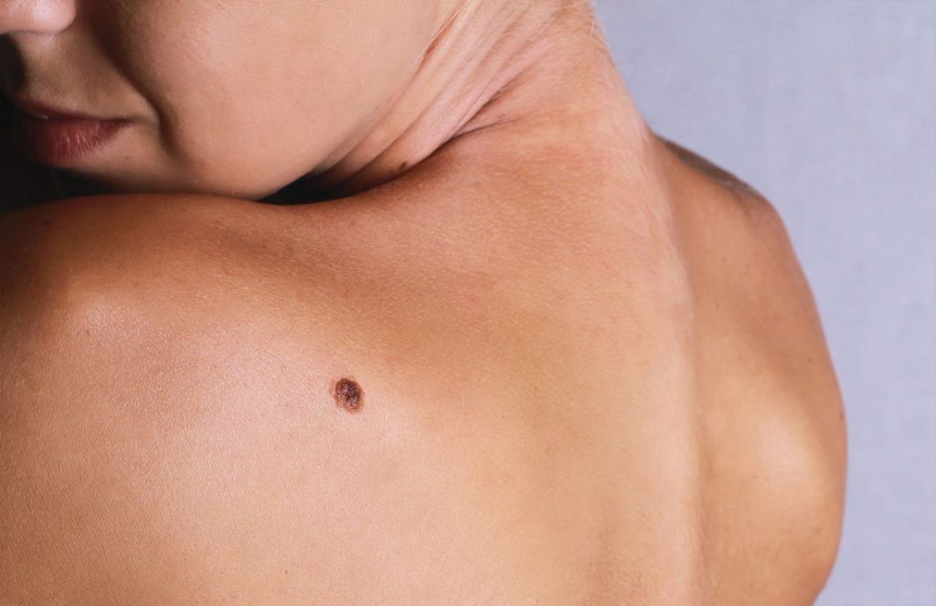
There are more to the diminutive, brown spots on our body than most of us know. Understanding and identifying the different types of moles is one of the best ways for early detection of skin malignancies.
Moles explained
Round, oval, flat or raised, a mole or nevus (plural – nevi) can occur on its own or in clusters on any part of our body. Moles comprise cells called melanocytes, which produce melanin in our skin. They are initially present in the epidermis (outermost layer of skin), dropping down to the dermis (middle layer) as the mole matures, growing more elevated and eventually losing its pigmentation as it reaches the final stage. Predominantly brown, moles can otherwise range from pinkish flesh tones to yellow, dark blue, or black. While moles may be present at birth, most of them do appear later in life, such as during pregnancy or puberty. However, moles rarely appear for the first time beyond the 50s — these should be treated with suspicion.
The precise causes of moles are yet to be fully understood. However, dysplastic nevi seemingly run in families. Exposure to the sun, particularly since childhood, and hormonal changes during adolescence are other postulated causes.
Types of moles
Every mole is unique to the point they have multi-factorial classifications — time of development, location in skin, and whether typical or atypical in appearance. Some of these classifications include:
Common (Typical) A common mole is identified by its distinct edges, smooth surface, and even pigmentation. Usually less than 6mm in diameter, common moles are typically found on skin regularly exposed to the sun. They range from flat to slightly elevated to the mature domed flesh-coloured mole. On rare occasions, these have the potential to turn into skin cancer.
Atypical A mole that is atypical in appearance may be a dysplastic nevus or even a form of pigmented skin cancer. Dysplastic nevi are usually larger than 6mm in diameter, asymmetrical, have hazy or irregular borders, and different shades of colour. They can be flat or raised. While most atypical moles are benign, it is important to have them examined by a qualified dermatologist to exclude pigmented skin cancers. Furthermore, a small proportion of dysplastic nevi may progress over years to melanoma (an aggressive form of skin cancer), so a person with many of them is at increased risk for melanoma.
Congenital Present at birth, congenital moles form in various sizes and are sometimes referred to as birthmarks. Medium-sized and large congenital nevi can potentially develop into melanoma later in life, and should be observed carefully as one enters adolescence and adulthood.
Acquired Most moles are acquired and turn up during childhood and early adulthood, especially with increased sun exposure. Although most are benign, sometimes they can turn cancerous with age.
Blue nevi Its colour is from the pigmented nevus cells being situated deeper in the skin than brown moles. They can emerge anywhere and are generally isolated. They range from light to dark blue, and are commonly small, round, raised or flat. Although blue nevi are benign, they may be difficult to differentiate from melanoma and may require surgical removal for testing.
Dangerous mole mimics
Melanoma The most dangerous form of skin cancer, melanoma is caused largely by intense, occasional ultraviolet exposure (most often via sun exposure or tanning beds) — especially in those who are genetically prone. Melanomas commonly arise de novo as atypical pigmented ‘moles’, while some develop from pre-existing moles. The majority of melanomas are black or brown, but can also be skin-coloured, pink, red, purple, blue or white. If detected and treated early, melanoma can be cured. Otherwise, it quickly spreads to other areas of the body, and turns fatal.
Basal cell carcinoma (BCC) The most common form of skin cancer in Singapore, BCCs may present as a pigmented lesion in Asian or darker skin types, or as red patches, pink growths, shiny bumps, non-healing sores or scar-like lesions. They typically appear in areas of high sun exposure such as face, neck and shoulders, often in sun-damaged skin with freckles and sunspots. BCCs usually grow locally, causing disfiguring ulcerated areas if not treated early.
Knowing if your mole is safe the ABCDE way

It’s crucial to take note of the moles on your body, and be alert to changes that occur over time, especially since certain types of moles are more prone to develop into skin cancer. For instance:
- There is a one in 20 risk that a large congenital melanocytic nevus may become cancerous, while smaller congenital nevi are less likely to turn cancerous
- Dysplastic nevi have an increased chance of developing into melanoma
The ABCDE method is a simple approach to noting changes. Consult a dermatologist if you notice the following signs:
A – Asymmetry If the two halves are not mirror images of each other, it is a warning sign of melanoma.
B – Border Benign moles have smooth, even borders, while the borders of melanoma tend to be uneven or ragged.
C – Colour Most benign moles come in a single shade, usually brown. Varying colours — shades of brown, tan or black — is a warning sign. A melanoma may also turn red, white or blue.
D – Diameter Moles larger than 6mm in diameter have a higher chance of being dysplastic or melanoma.
E – Evolution Benign moles appear the same over long periods of time. Be cautious when a mole starts to change in any way in a short period, including looking unusual, changing in size, shape or colour, and starting to itch, bleed, ooze or ulcerate. When a mole starts to evolve, consult a dermatologist.


