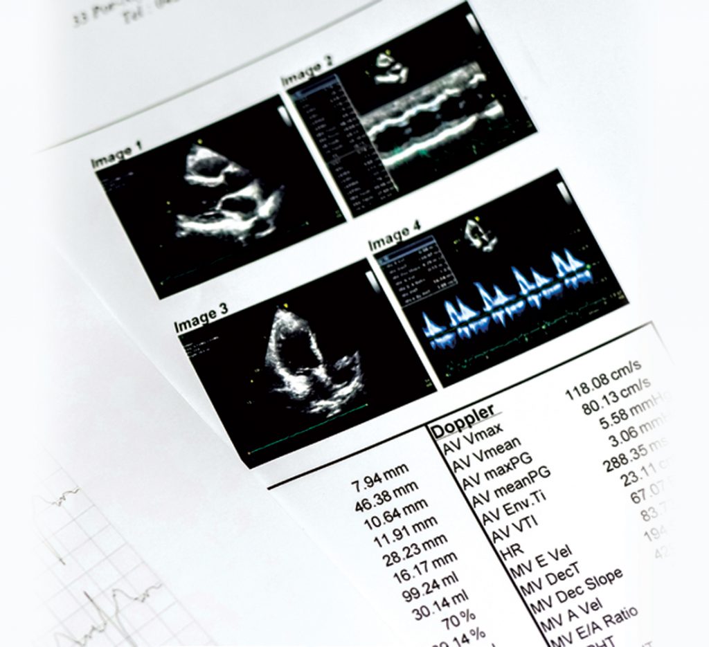
ECHOCARDIOGRAM uses ultrasound to produce detailed images of the heart’s structure and function.
During a transthoracic echocardiogram A transducer pressed against your chest sends ultrasonic waves to the heart. Echoes inside the heart are recorded and converted into images on a monitor. A contrast agent may be injected through an intravenous (IV) line to make the heart structure more distinct.
What can echocardiograms assess?
- Heart size (enlarged heart chambers, thickened heart wall)
- Pumping strength (ejection fraction, cardiac output)
- Speed and direction of blood flow
- Valve malfunctions (inadequate blood flow, blood leakage)
- Heart defects (abnormal connections between the heart and major blood vessels, congenital heart defects)
CARDIAC COMPUTERISED TOMOGRAPHY (CT) SCAN is a non-invasive imaging procedure. Instead of catheters, it uses a powerful X-ray machine to visualise your cardiac anatomy, coronary circulation, and blood vessels. Advances in cardiac CT have accelerated in the past few years. With the use of multi-detector CT systems, coronary CT angiography has been shown to have high negative predictive value for ruling out coronary artery disease (CAD).
During a cardiac CT scan Lay still on a table inside the doughnut-shaped machine as an X-ray tube rapidly moves around your chest to collect images of your heart from different angles. The contrast agent is injected through an IV line in your arm.
What can cardiac CT scans assess?
- Heart muscle
- Coronary arteries
- Pulmonary veins
- Thoracic and abdominal aorta
- Pericardium (sac enclosing the heart)
MYOCARDIAL PERFUSION IMAGING (MPI) is a non-invasive test that allows doctors to assess the function and blood flow of the heart at stress and at rest.
During an MPI
A radioactive tracer is injected and takes images of the heart using a gamma camera or Positron Emmision Tomography (PET) camera. MPI is indicated for the diagnosis of patients with suspected CAD and risk stratification of patients with known CAD, when they are unable to exercise, with abnormal baseline electrocardiogram (ECG), heavily calcified coronary arteries, allergy to contrast or an equivocal cardiac screening test.
What can MPI assess?
- Assess coronary vasodilator flow reserve, progression of coronary artery disease, and stratify risk of future cardiovascular outcomes
CARDIAC CATHETERISATION can diagnose and treat heart and blood vessel conditions. A common procedure is coronary angiogram.
During a cardiac catheterisation
A tube (sheath) is inserted into an artery in the groin or arm. A hollow, flexible and longer tube (guide catheter) is then inserted into the sheath. The guide catheter is threaded through that artery until it reaches the heart. A dye that makes blood vessels visible on X-ray is injected, while the X-ray machine takes a series of images. If necessary, angioplasty or stenting can be done to open clogged arteries.
What can cardiac catheterisations assess?
- Blockage in blood vessels that cause chest pain or put you at risk of heart attack
- Pumping function


