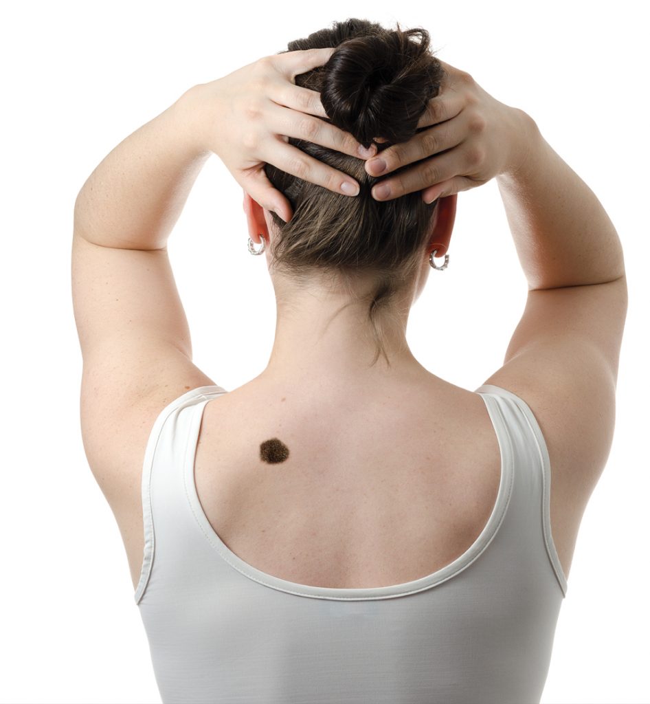
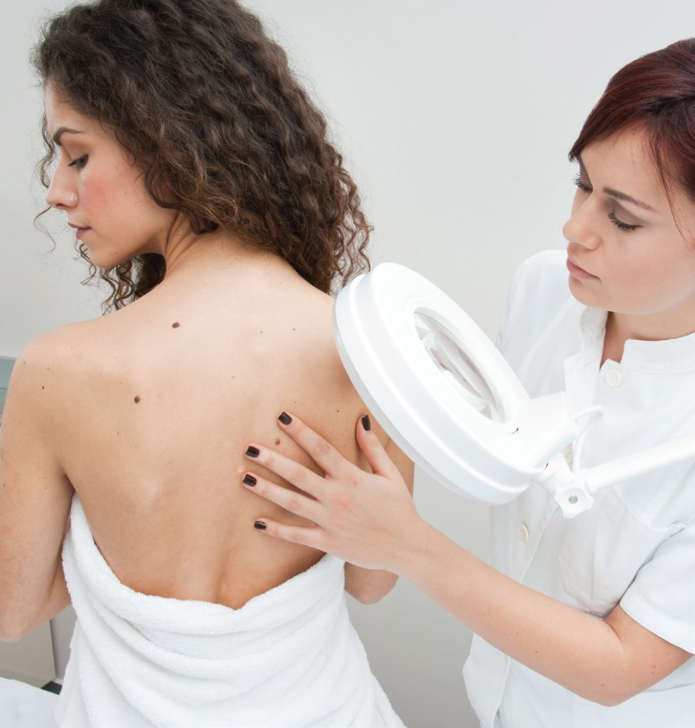
Regular self-examination may help stop skin cancer in its tracks.

Melanoma is an aggressive form of skin cancer that can invade nearby tissue and spread to other parts of the body. It develops in the cells that produce melanin – the pigment that gives skin its colour.
Normally, cells develop in a controlled and orderly way – healthy new cells push older cells toward the skin’s surface, where they die and eventually fall off. But when some cells develop DNA damage, new cells may begin to grow out of control and eventually, form a mass of cancerous cells. Skin cancer can be treated successfully, especially when it is in the ‘in-situ’ stage, or hasn’t invaded deeper tissues.
Melanoma can arise from existing moles or in previously normal-looking skin and can appear anywhere on your body. It can also form in your eyes and rarely, in internal organs, such as the intestines. It appears to be increasing in those under 40, and especially in women. Knowing the warning signs of skin cancer can help ensure that cancerous changes are detected before the disease has spread. Consult a doctor if a pre-existing mole or a newly-appeared pigmented spot displays the following ABCD criteria:
A: Asymmetry. The shape of one half is not the mirror image of the other
B: Border irregularity. The edges are irregular, scalloped, notched or blurred
C: Colour irregularity. Normal moles are generally a uniform colour such as tan, brown or black but suspicious-looking ones are those that have many shades or an uneven distribution of colour
D: Diameter. Increasing, or more than 6mm E: Evolving. Changes in size, colour, thickness, shape and texture, swelling, redness, hardening, itching, bleeding with no injury, especially over a few weeks or months
Cancerous moles vary greatly in appearance. Some may show all the changes listed above, while others may have only one or two unusual characteristics.
Risk factors
Those with a family or past history of melanoma are at higher risk of developing the condition and should be especially careful about regular skin self-examination as well as regular checks by a dermatologist as often as three, six or 12 months depending on individual risk factors.
Other risk factors for melanoma are:
- Having many moles (more than 50)
- Having more than five dysplastic nevi (atypical moles) is associated with 10 times increased chance of melanoma – up to 10% of dysplastic nevi may develop into melanoma
- Repeated sunburn or one episode of severe blistering sunburn
- High lifelong sun exposure (eg. those who work outdoors)
- Previous use of sunlamps or tanning booths or frequent suntanning
- Skin that burns easily (usually fair skin with freckles, fair or red-haired, blue- or grey-eyed)
- Large congenital nevi (present at birth), especially those larger than the size of a palm
Sun-damaged areas are more prone to skin cancer. However, melanoma may also affect non-exposed areas such as the scalp and genitalia. Darker skinned people are at lower risk of melanoma but when they develop it, it tends to affect non-exposed areas such as the palms, soles, and around fingers and toenails. These areas should be included in a complete skin examination, preferably done monthly.
Doing a self-examination
The best time for a skin examination is after a shower. Use a full-length mirror and a handheld mirror to examine all parts of your skin in a bright room.
Move systematically from head to toe, starting with the face, neck, scalp, ears and behind the ears. A hairdryer or comb may be used to move hair out of the way to examine the scalp.
Examine the front and back of the body, lift arms to examine the sides and underarms.
Examine the arms next, including the undersides, fingernails and palms.
Examine the front and back of legs, including the buttocks and genitalia.
Sit down and examine the soles, toes, web spaces and toenails.
If you have too many moles to remember, it is helpful to take photographs at regular intervals so that any new lesions can be more easily detected. Your dermatologist can also help with ‘whole body photography’.
Look for:
- New moles that appear different from your other moles
- Changes in shape, size, colour, texture of existing moles
- A new red or darker coloured flaky patch that may feel a little raised
- A sore that doesn’t heal within a few weeks
- A new skin-coloured firm bump
Taking action
If you detect suspicious lesions, take a photograph and schedule a consultation with a dermatologist, who will observe for changes since the last photo, examine them carefully with a magnifying instrument or a dermatoscope, and may recommend serial photography of atypical moles to observe for changes, or a skin biopsy to determine if there is skin cancer.
While it is common to have new moles when you are in your 20s and 30s, moles that start appearing in those who are in their 50s onwards should be viewed with suspicion as they could indicate melanoma or other forms of pigmented skin cancer. A dermatologist should be consulted in such situations.

DID YOU KNOW?
Skin cancer (including melanoma) is ranked the 6th and 7th most common cancer in Singapore men and women respectively, with about 3,000 new cases a year. Although melanoma accounts for less than 2% of skin cancer worldwide, it causes the most deaths.


